The reason why earthworms will not die if cut into two sections
Earthworms themselves are eukaryotic cell organisms
To be precise, it does not necessarily grow into two. Earthworms are a higher type of annelid. The structure is more complicated than many annelids. The digestive system and circulatory system appear, which is the prerequisite for the development of vertebrates!
It is also distributed with dorsal blood vessels. If it loses too much blood when cutting, it may not become two. There are many external factors that can affect its growth. If it is under temperature, pH and sterilization conditions, it may survive. Coming down!
It is like tissue culture of plants.Of course, cutting off earthworms and tissue culture is not a principle, but the lower the organism can be expressed, the greater its ability to develop into the same independent individual.
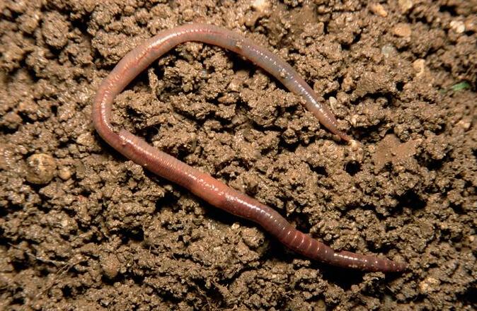
Earthworms are a kind of lower annelids, born with a head and tail, mouth, stomach, intestines, and anus. Their bodies are made up of two pointed tubes set together. The outer layer is a body wall connected ring by ring, in which there are many muscular systems composed of mesoderm cells. There is a digestive tract in the body, which runs through the layers of the diaphragm from beginning to end. Between the inner and outer tubes, it is filled with body cavity fluid, and there is a small hole on the ventral surface of each diaphragm, which becomes a channel for body cavity fluid to pass through the body.
If an earthworm is cut into two sections, the muscle tissue on the section will immediately strengthen its contraction, and part of the muscle tissue will quickly dissolve to form new cell clusters. At this time, the white blood cells in the blood gather on the cut surface at the same time, forming a special embolism, which makes the wound close quickly. Its mesodermal cells are very strong.
Environmental requirements for the growth of earthworms
1. Temperature
Generally speaking, the active temperature of earthworms is 5-30℃, 0-5℃ enters a dormant state, and they die below 0℃, and the optimum temperature is about 20-27℃, which is also the optimum temperature for earthworm cocoon eggs, above 32℃ Stop growing and die above 40℃. Therefore, earthworm breeding: shade sheds should be built in summer and autumn to cool down (mulberry gardens have natural shading conditions, but mulberry trees need to be covered with shade temporarily after summer felling and before germination and growth), and the shed should be heated up in winter (You can use the equipment already in the silkworm house) or mulberry orchard with wheat-covered straw to keep warm, so as to facilitate the normal growth and reproduction of earthworms.
2. Humidity
Earthworms use their skin to breathe, so their body must be kept moist. The water in the body of the earthworm accounts for more than 75% of its body weight. Preventing the loss of specific water is the key to the survival of the earthworm. Therefore, the humidity of the feed should be kept at about 70%.
3. Acidity (pH value)
Earthworms grow better in the pH range of 6-8, and produce the most cocoons.
4. Ventilation
Earthworms breathe by oxygen diffused into the soil. The better the aeration of the soil, the more vigorous its metabolism, which not only produces more cocoons, but also shortens the maturity period.
5. Food
Insufficient or low-quality feeding will cause maggots to fight for food between calls, resulting in decreased fertility, spread of pests and diseases, increased mortality, and some earthworms escape or grow slowly.
Physiological mechanism of earthworms
Body wall, secondary body cavity
The body wall of an earthworm is composed of corneum, epithelium, circular muscle layer, longitudinal muscle layer and body cavity epithelium. The outermost layer is a single layer of columnar epithelial cells, and the secretions of these cells form a cuticle. This membrane is extremely thin, composed of collagen fibers and non-fiber layers, with small holes. Columnar epithelial cells are mixed with glands
Cells, divided into mucous cells and protein cells, can secrete mucus to moisten the body surface. When earthworms encounter severe irritation, a large amount of mucus cells secrete and wrap the body into a mucous membrane, which has a protective effect. There are short basal cells at the base of epithelial cells, which some people think can develop into columnar epithelial cells. Sensory cells gather to form sensory organs, which are scattered among the epithelial cells, and the base is connected with the nerve fibers of a thin layer of nerve tissue under the epithelium. In addition, there are photoreceptor cells, located at the base of the epithelium, and also connected to the nerve fibers underneath.
The muscles of earthworms belong to the oblique muscle, which generally occupies about 40% of the body’s volume. The muscles are strong and flexible. The longitudinal muscle layer of some body segments of the earthworm contracts and the circular muscle layer relaxes, then this segment of the body segment becomes thicker and shorter, and the bristles that are born on the body wall obliquely extend and insert into the surrounding soil; at this time, the previous segment of the body segment The circular muscle layer contracts and the longitudinal muscle layer relaxes. This segment of the body segment becomes thinner and longer, and the bristles retract to disengage from the surrounding soil pendulum. In this way, the bristles of the latter segment of the body segment push the body forward. In this way, the muscle contraction waves are gradually transmitted from front to back along the longitudinal axis of the body.
The body cavity is divided into a plurality of coelomic compartments by a septum according to the body segments, and each compartment is communicated with a small hole. Each integrated cavity is formed by the development of left and right body cavities. The outer side of the body cavity sac forms the wall body lumen membrane, the inner side forms the visceral body lumen membrane except for most of the middle, and the dorsal and ventral sides form the dorsal mesenteric and abdominal mesenteric. The ventral mesentery of earthworms degenerates, and only the part between the intestine and abdominal blood vessels exists; the dorsal mesenteric has disappeared. The part between the front and rear body cavity sacs is tightly attached to form a septum. Some species have no diaphragm in the esophagus.
Digestive system
The digestive tube runs longitudinally in the center of the body cavity, passing through the diaphragm, and the muscular layer of the tube wall is developed, which can enhance peristalsis and digestion. The digestive tract is divided into parts such as mouth, mouth, throat, esophagus, gizzard, stomach, intestine, and anus. The mouth can be turned out to take food. The pharyngeal muscles are well developed, the muscles are contracted, and the pharyngeal cavity is enlarged to aid in feeding. There are single-cell pharyngeal glands outside the pharynx, which can secrete mucus and proteases, moisturize food and initially digest.
The short and thin esophagus after the pharynx has esophageal glands on its wall, which can secrete calcium and neutralize acidic substances. Behind the esophagus is a muscular gizzard, lined with a thick keratinous membrane, which can grind food. The ectoderm forms from the mouth to the gizzard and belongs to the foregut. The digestive tube behind the gizzard is rich in capillaries and multiple glands, called the stomach.
There is a ring of gastric glands in front of the stomach, which functions like pharyngeal glands. After the stomach starts from the 15th body segment, the digestive tube expands to form the intestine, and its dorsal center is recessed into a blind tract (typhlosole), which increases the area of digestion and absorption. The digestion and absorption functions are mainly carried out in the intestine. The visceral lumen membrane in the outermost layer of the intestinal wall is specialized into yellow cells. From the 26th somite, a pair of cone-shaped cecum (caeca) extends forward on both sides of the intestine, which secrete a variety of enzymes and are important digestive glands.
The stomach and intestine are derived from the endoderm and belong to the midgut. The hindgut is short, occupying more than 20 body segments at the rear end of the digestive tract, with no blind tract and no digestive function. The anus opens outside the body. The digestive system of earthworms is composed of more developed digestive tracts and digestive glands. The digestive tract is composed of mouth, pharynx, esophagus, crop, gizzard, stomach, small intestine, cecum, rectum and anus.
Circulatory system
The earthworm is very special. Just like its body is divided into sections without obvious merging, its heart is also divided into several sections along the front of the body, usually 4 to 5, in a ring shape, like enlarged blood vessels, so There are also ring vessels. The back of the ring-shaped heart is connected to the back-to-front dorsal blood vessels, and the ventral surface is connected to the front-to-back abdominal blood vessels. The abdominal blood vessels have branches connected to the subneural blood vessels from front to back. Compared with blood vessels, the annular heart has a thicker muscle wall and can pulsate, and there are valves that open in one direction to ensure blood flow from the dorsal blood vessels to the abdominal blood vessels. Generally speaking, the pulsation of these independent ring-shaped hearts powers the blood flow. The direction of blood flow is from back to front (in the dorsal blood vessel), from back to the abdomen (ring heart), and from front to back. (Abdominal blood vessels and subnervous blood vessels).
Respiration and excretion
The excretion organ of earthworms is the metanephric duct. In general, each body segment has a pair of typical metanephric ducts, which are called great renal ducts. Earthworms of the genus Cyclophora do not have a large renal tube, but have three types of small tube: the small body wall (parietal micronephridium) is located on the inner surface of the body wall and is extremely small. Each body saves 200 to 250. There is no renal orifice at the inner end, and renal foramen. Open on the body surface. Septal micronephridiumm (septal micronephridiumm) is located on the front and back sides of each diaphragm after the 14th body segment. Generally, there are 40 to 50 on each side. There is a funnel-shaped renal orifice, with cilia, and a small renal tube with cilia in the lower part of the internal organs. The excretion tube without ciliated lumen opens into the intestine. The pharyngeal micronephridium (pharyngeal micronephridium) is located at the pharynx and on both sides of the esophagus, without a renal orifice, and opens to the pharynx. The latter two types of renal tubes are also called digestive renal tubes. Various types of small renal tubes are rich in capillaries, and some of the kidney orifices open in the body cavity, so it can eliminate the metabolites in the blood and in the body cavity. Yellow cells outside the intestine can absorb metabolites, then fall off the body cavity fluid, enter the kidney orifice, and be discharged from the kidney tube.
nervous system
Earthworms are typical cable nerves. The central nervous system has a pair of suprapharyngeal ganglia (brain) located on the dorsal side of the third body segment and hypopharyngeal ganglia located on the ventral side of the third and fourth body segments. A ventral nerve cord extending from the pharyngeal ganglion to the back of the body, with a ganglion in each section). In the peripheral nervous system, there are 8-10 pairs of nerves from the anterior side of the suprapharyngeal ganglion, which are distributed to the anterior mouth, oral cavity, etc.; the hypopharyngeal ganglion divides the nerves to the body wall of several somites in the front of the body. Each ganglion of the abdominal nerve cord sends out 3 pairs of nerves distributed in the body wall and various organs. The nerves extending from the suprapharyngeal ganglion to the digestive tract are called the sympathetic nerve system.
Every nerve in the peripheral nervous system contains sensory fibers and motor fibers, which have conduction and response functions. Sensory nerve cells can transmit the stimulus received by the epithelium to the adjustor neuron of abdominal neuron, and then transmit the impulse to the motor nerve cell, connect to the muscle and other reactors through the nerve fiber, and cause the reaction. This is simple Reflex arc. The three giant fibers in the abdominal nerve cord run through the entire cord and transmit impulses extremely fast, so earthworms respond quickly to stimulation.
Reproductive system
Female reproductive organs: 1 pair of ovaries, very small, composed of many very thin ovarian tubes, located on the back side of the diaphragm before the 13th somite, and a pair of oviduct funnels, located on the front side of the diaphragm after the 13th somite. Followed by a short oviduct. The two fallopian tubes meet under the ventral nerve cord of the 14th somite and open at the midline of the abdomen, called the female genital foramen. There are also 3 pairs of seminal receptacle (4 pairs for P. differingens, 2 pairs for P. aspergillum and P. californica), which ranks in the 7.8.9 somite. The seminal vesicle is composed of an ampulaa, a tube and a diverticulum. It is a place to store sperm. On both sides of the ventral surface between the 6/7.7/8.8/9 somites of the seminal vesicle hole.
Male reproductive organs: 2 pairs of testes, very small, located in the seminal sac (seminal sac) of the 10th and 11th somite, 2 pairs of seminal funnels, just below the testis, enlarged front end, mouth with cilia, followed by thin vas deferens . The two tubes are combined into one in the 13th body segment, extending backwards, opening on both sides of the 18th body segment, which are male genital holes. A pair of prostate gland, located on one side of the male genital opening, the prostate tube opens at the end of the vas deferens, and the secretion of mucus waves is related to the movement and nutrition of sperm. The seminal vesicles are connected to the seminal vesicles in the 11th and 12th body segments. ) Are connected, and the seminal vesicles are filled with nutrient solution. After the spermary produces sperm cells, it first enters the seminal vesicle to develop, and when the sperm is formed, it returns to the testis sac and is output from the vas deferens through the seminal funnel.