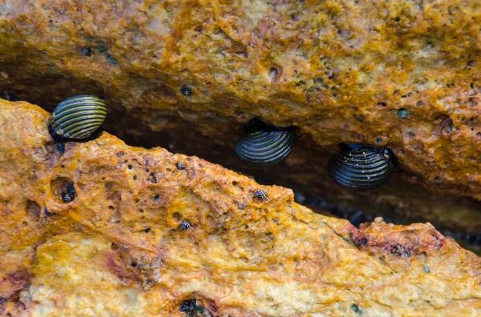
The reason why some shellfish like to live in rocks
If children have the opportunity to explore treasures in the sea, they will definitely find that there are many holes in some hard stones, and some shellfish such as stone clams and sea shoots live in the caves. It turns out that these holes were chiseled by some shellfish such as stone clams and sea bamboo shoots. So, why do they spend so much effort to dig these holes? It is very strange to say that their growth and development cannot be separated from stones. If they only eat food without chiseling rocks, they will not grow up.
Only when they continue to drill through the stone can they grow and develop. The stones are so hard, how do they make holes? It turns out that their feet can secrete an acidic liquid that can corrode the rock and make the rock soft, and then stand on the corroded rock with your feet and foot tubes Supporting the body, making the shells spin quickly, the teeth on the shells are like saws, constantly rubbing the stone surface, drilling holes. The 147 stones of the seasun are bigger. Because it reproduces fast and abundantly, and likes to live together in groups, many rocks are drilled like honeycombs by them. Their rock-cutting ability often destroys some buildings in ports and docks. Therefore, in order to prevent them from destroying, port buildings use more granite that they cannot drill.
Physiological characteristics of shellfish
Digestion: In the original deposit-feeding type, the digestion of food and the structure of the digestive tract still maintain the shape and function of the original mollusk. For example, the stomach wall of the protobranch is very thin, and the structure of the stomach and crystal rods is still retained. Food is digested extracellularly in the stomach, and digested and absorbed intracellularly in the digestive caecum. There are no tentacles around the mouth of the digestive tract in advanced filter feeding species. The upper stomach wall and the screening area are underdeveloped, while the crystal rod capsule is developed and protrudes beyond the stomach wall. The mucus secretions in the capsule are solidified to form a crystal rod, and digestive enzymes (amylase, lipase) are adsorbed on the crystal rod. The cilia in the capsule wall cause the crystal rod to rotate continuously, and the top of the crystal rod is dissolved to release digestive enzymes. For extracellular digestion, the rotation of the crystal rod also plays a role in mixing food and enzymes. The top of the crystal rod is constantly worn by food, and the back end can be constantly replenished. The rotation of the crystal rod also allows tiny food particles to enter the gastric caecum for intracellular digestion and absorption. Indigestible food residues are discharged out of the body through the intestines, anus, and water outlets. A slow but continuous flow of food in the digestive tract is unique to the lobular and branchial filter feeders. Gills are carnivorous animals. The muscular stomach wall is surrounded by chitin to form a pulverized stomach. Its crystal rods are underdeveloped and extend into the stomach in the form of small sticks with more developed digestive enzymes.
Circulation and gas exchange: All bivalves have open circulation. The pericardial cavity is located on the back of the body. There is one ventricle and two atrial appendages in the pericardial cavity. There are valves between the ventricles and the atrial appendages to prevent blood reflux. In the protobranch and silk branch, only the anterior aorta passes forward from the ventricle, such as mussels, and in the valve branch, in addition to the anterior aorta, the posterior aorta passes back from the ventricle, such as mussels. After the blood flows out of the arteries, it branches to the front end of the body, feet, internal organs, etc., and forms sinusoids in the tissues. After the sinuses, it merges into blood vessels, passes through the kidneys and gills, and then flows back to the auricles and ventricles. The gill is its main gas exchange place. When the water passes by, the amount of oxygen taken up by the gill is less than that of other mollusks. This may be related to the larger surface area of the gills than other molluscs. In addition, all bivalves have more or less developed mantle circulation. After the blood flows out from the arteries, it directly flows into the mantle to form sinusoids. After the sinusoids merge into blood vessels, they flow directly back to the atrial appendage or pass through the kidneys. After the metabolites are discharged, they flow back to the atrial appendage. The jacket circulation is also an auxiliary place for gas exchange. Respiratory pigment does not exist in the blood of most bivalves, and only a few species such as cockles and Lima have heme, which makes the mantle and other tissues appear red. The septal gills have disappeared, and the gas exchange is completely carried out by the mantle.
Excretion: The excretion system of bivalves is a pair of metanephros, located on the ventral surface of the pericardial cavity. The kidney is in a long tubular shape, but the inner renal orifice opens at the front end of the pericardial cavity, and the outer renal orifice opens below the inner renal orifice of the outflow, so the rear end of the kidney is folded back, and there is a gland called the gland in the front half of the kidney. This performs waste filtering, followed by the bladder part, which is the storage place for metabolites. There is no distinction between the glandular part and the bladder part of the protobranch kidney. The kidneys of freshwater species have a reabsorption of salt, so the excreted urine is hypotonic.
Nerves and sense organs: The nervous system of the bivalves is relatively simple. The original species has four pairs of ganglia, brain, side, foot, and victory. The more evolved species, the brain and the lateral ganglia merge, so there are only 3 pairs of ganglia, the brain The lateral ganglia are located on both sides of the esophagus. They control the anterior adductor muscle and coordinate the movement of the feet and shells. The visceral ganglion is located on the posterior adductor muscle column. It controls the contraction of the internal organs and the posterior adductor muscle. The foot ganglion is located in the front muscle of the foot and controls the movement of the foot. In addition, it is located between the lateral cranial ganglion and the visceral ganglia. There are two pairs of nerve cords connecting the ganglia between the lateral ganglia and the cerebropedal ganglia. The sense organs are underdeveloped. In some species with greater mobility, such as scallops, there are pairs of small tentacles on the middle fold of the mantle limbus, which contain tactile and chemical sensory cells, and many ommatidiums. Developed, even including crystals and omentum, can feel the change of light intensity. In addition, there is a pair of balance capsules around the foot ganglia to control the balance of the body. Many species have some sensory epithelium under the posterior adductor muscle or around the water outlet, called sniffer, which can sense the changes in water quality and flow.